
Labral tear of the hip
A hip labral tear affects the ring of cartilage around the edge of the hip socket. It may cause groin pain, clicking, catching or discomfort with sitting, twisting and exercise. Many tears can be managed with physiotherapy and activity changes.
At the Specialist Orthopaedic Surgery Clinic in East Melbourne, orthopaedic review, physiotherapy, myotherapy and strength testing work together in one place, so your care can be matched to your hip and the activities you want to return to.

What is a hip labral tear?
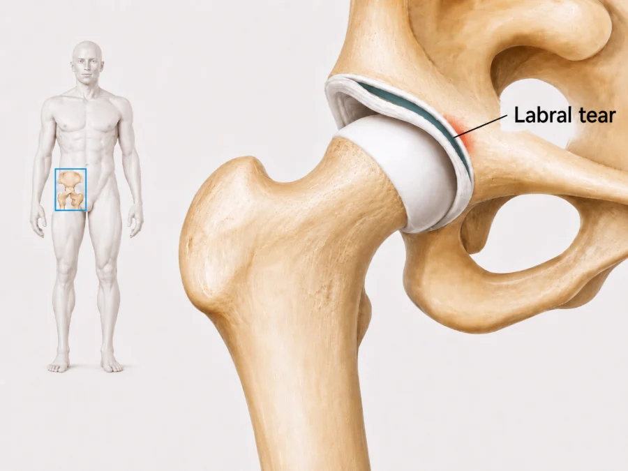
The labrum is a ring of strong, flexible cartilage attached to the rim of the hip socket. It deepens the socket, supports stability and helps seal the joint so it moves smoothly.
A labral tear is a split or separation in this tissue. It can follow an injury, develop gradually with repeated loading, or occur alongside hip impingement, dysplasia or arthritis. Labral changes are also common on scans in people without pain, so the finding is always weighed against your symptoms and examination.
Does this sound like you?
Arrange an assessment if groin pain, painful clicking or catching continues, limits work or sport, or has not improved with a structured rehabilitation program. Review is especially useful if the hip keeps locking or giving way.
How it is diagnosed
Diagnosis starts with the pattern of groin pain, the movements that bring it on and whether there is clicking or catching. Hip movement and strength are examined, and other possible sources of pain such as the lower back and nearby tendons are checked.
MRI, sometimes with contrast placed into the joint, may show the tear and any cartilage changes. Because labral changes can be present without pain, the scan is only useful when it matches your symptoms and examination.
Treatment and recovery
Treatment depends on whether the tear is the main source of pain, the shape of the hip and the health of the nearby cartilage. Initial care usually includes activity changes, physiotherapy and a gradual return to loading.
First weeks
Settle the pain
Movements that repeatedly pinch the hip are adjusted while general fitness is kept up. Short-term pain relief can be discussed with your GP.
Physiotherapy
Build control
Exercises build hip and trunk strength and movement control. Myotherapy may ease tight muscles but does not repair the labrum.
Building up
Return to activity
Load is increased gradually for work, exercise and sport. Strength testing can track readiness for higher load activity.
If progress stalls
Review the plan
If pain or catching remains limiting despite a structured program, an injection or a surgical opinion may be arranged. Injections are not given at the clinic. If one is recommended, your GP can arrange it, usually through a radiology practice.
Exercises you may be given
Your physiotherapist will choose the right ones and the right level for you.
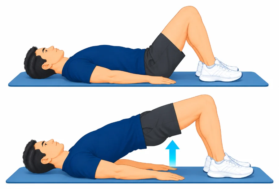
Bridge
Lie on your back with knees bent and feet flat. Squeeze your buttocks and lift your hips until your body is in a straight line, then lower slowly.
2 to 3 sets of 10
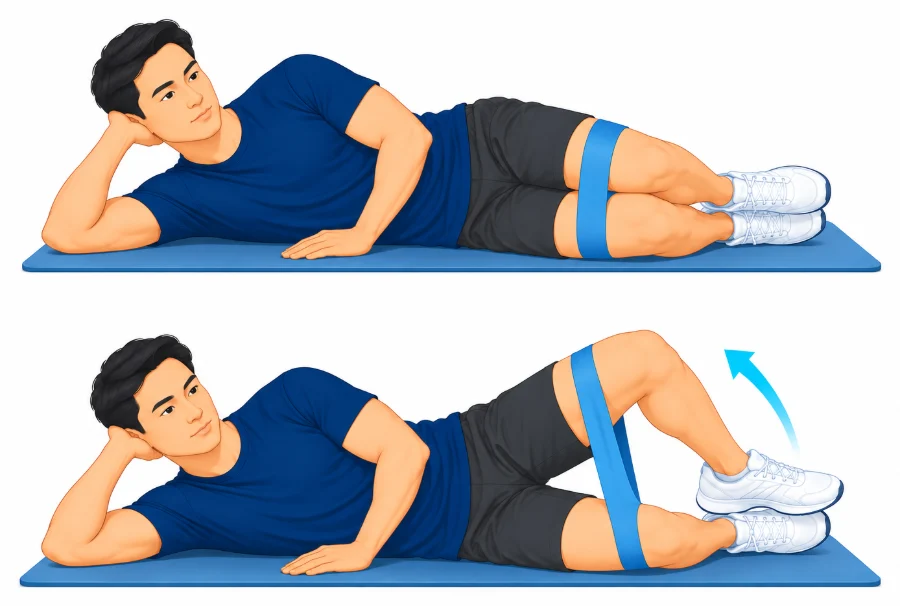
Clam
Lie on your good side with hips and knees bent. Keep your feet together and lift the top knee without rolling your pelvis back, then lower.
2 to 3 sets of 10 to 15
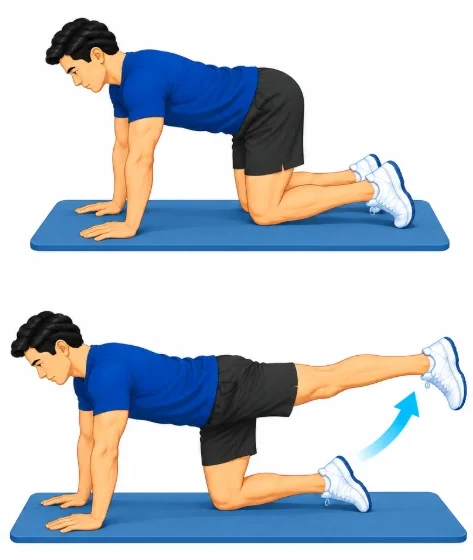
Four-point leg reach
Kneel on hands and knees with a flat back. Slowly slide one leg straight out behind you and lift it to hip height without twisting, then return and swap.
2 sets of 8 each side
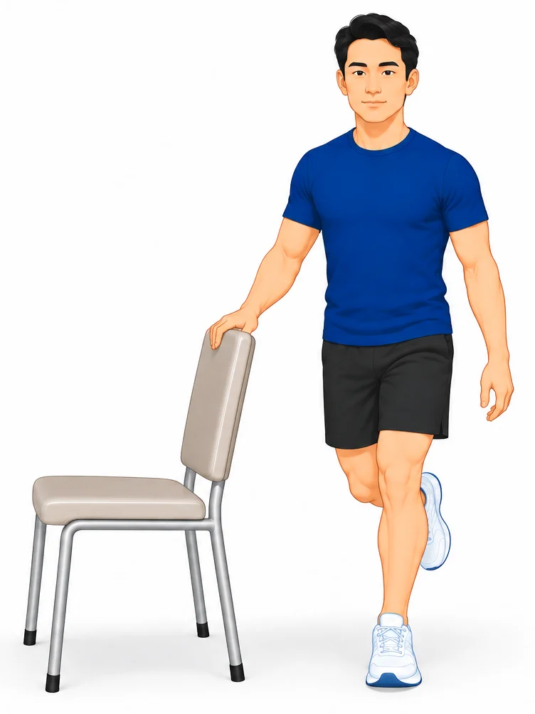
Single-leg balance
Stand tall on the sore leg near a bench for support. Keep your pelvis level and your knee soft, and hold your balance.
3 to 5 holds of 30 seconds
Other treatment options
Injections
An image-guided hip joint injection may give temporary relief and can help show how much pain comes from inside the joint. A good response does not guarantee that surgery will help.
When surgery is needed
Hip arthroscopy may be considered when pain, catching or loss of function remains substantial despite appropriate rehabilitation, and the tear is judged to be a meaningful source of symptoms without advanced arthritis. A tear seen on MRI without matching symptoms is not a reason for surgery. Prof Phong Tran performs hip arthroscopy and labral repair, and his website explains the options and recovery.
Common questions
Does every hip labral tear cause pain?
No. Labral changes often appear on MRI in people without symptoms. The scan must match the pain pattern, examination and hip shape.
Can symptoms improve without surgery?
Yes. Activity changes and physiotherapy can improve strength, movement control and tolerance for daily activity and sport.
Do I need an MRI arthrogram?
Not always. A standard MRI may be enough. More detailed imaging is used only when it is likely to change treatment.
When is hip arthroscopy considered?
It may be discussed when symptoms remain limiting despite rehabilitation and a treatable labral or impingement problem is present without advanced arthritis.
Can I return to sport after labral repair?
Many people do, but timing varies. Rehabilitation often takes four to six months or longer and depends on cartilage health, strength and the demands of your sport.
Book an assessment
An assessment can help work out whether a labral tear is behind your symptoms and which treatment may help. Physiotherapy with A/Prof Amir Takla and Imogen Clark, myotherapy with Joanne VanDer Haar, and specialist review with Prof Phong Tran, all at 166 Gipps Street, East Melbourne. Please bring any previous hip imaging and details of rehabilitation already tried.