
Osteonecrosis of the hip (avascular necrosis)
Osteonecrosis of the hip, also called avascular necrosis, happens when the blood supply to part of the ball of the hip is reduced. This can weaken the bone and, in some cases, cause the joint surface to collapse. Treatment depends on the stage of the condition, the amount of joint damage and how much the symptoms affect you.
At the Specialist Orthopaedic Surgery Clinic in East Melbourne, orthopaedic review, physiotherapy and strength testing are coordinated in the same rooms, so your hip can be monitored and supported as one plan.

What is osteonecrosis of the hip?
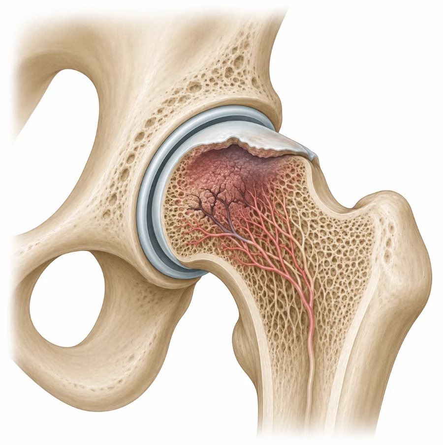
The ball at the top of the thigh bone, called the femoral head, needs a steady blood supply to stay healthy. In osteonecrosis part of this bone loses its blood supply and gradually weakens.
Over time the weakened bone may lose its normal rounded shape. If the surface collapses, the cartilage covering the joint can be damaged and arthritis may develop. One or both hips can be affected, and the stage of the bone changes helps decide which treatments may suit you.
Does this sound like you?
Arrange an assessment if deep groin or buttock pain is persistent, worsening, or affecting sleep or walking, especially if you take corticosteroid medicines or have another known risk factor. Osteonecrosis is easier to manage before the ball collapses, so ongoing unexplained hip pain should not be ignored.
How it is diagnosed
Diagnosis starts with the pattern of pain and a review of possible risk factors, including past hip injury, corticosteroid treatment, alcohol use and medical conditions. Hip movement, walking and function are examined.
MRI is the most useful test when osteonecrosis is suspected, as it can show early bone changes before they appear on X-ray and how much of the ball is involved. Blood tests are occasionally arranged to look for an underlying medical cause.
Treatment and recovery
Treatment is guided by the size and position of the affected area, whether the ball has started to collapse and how much pain you have. Regular review is often advised because the condition can progress even when symptoms are mild.
First steps
Protect the hip
High-impact activity and heavy loading are reduced. A stick or crutches may ease pressure through a painful hip for a time.
Risk factors
Address the cause
Alcohol intake and corticosteroid use are reviewed where possible, together with your GP or treating doctor.
Physiotherapy
Keep moving
Gentle exercise keeps comfortable movement, strength and fitness without repeatedly flaring the hip. Myotherapy may ease nearby muscles.
Ongoing
Monitor and review
Regular review and repeat imaging track any change. A surgical opinion may be arranged if the condition progresses or pain remains significant.
Exercises you may be given
Your physiotherapist will choose the right ones and the right level for you.
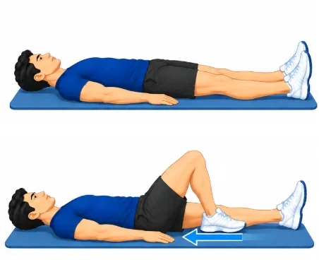
Heel slides
Lie on your back with legs straight. Slowly slide the heel of the sore leg towards you, bending the hip and knee as far as is comfortable, then slide it back.
10 slow repeats, twice a day
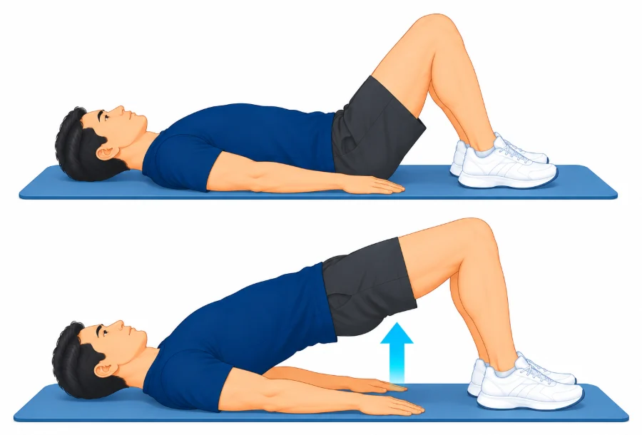
Bridge
Lie on your back with knees bent and feet flat. Squeeze your buttocks and lift your hips a comfortable height, then lower slowly.
2 sets of 10
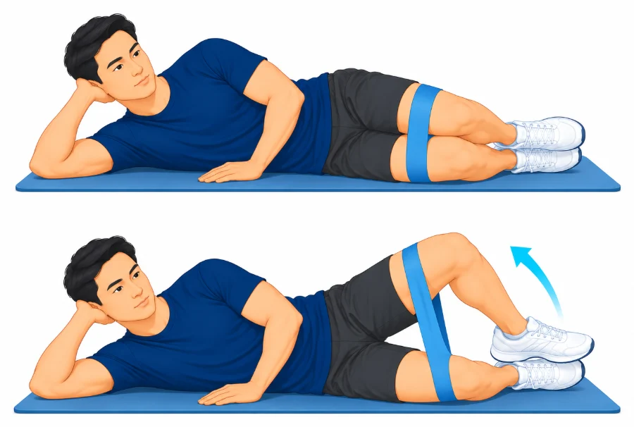
Clam
Lie on your good side with hips and knees bent. Keep your feet together and lift the top knee without rolling your pelvis back, then lower.
2 sets of 10
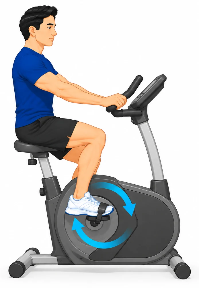
Exercise bike
Ride a stationary bike with the seat set high enough that the hip does not bend too far. Keep the resistance light and the pace easy.
10 to 15 minutes, easy pace
Other treatment options
Pain relief and walking aids
Pain relief should be reviewed with your GP, taking your other health conditions and medicines into account. A walking stick or crutches can reduce load on the hip while it is monitored. These steps ease symptoms but do not restore the blood supply to the bone.
When surgery is needed
Before the ball collapses, a joint-preserving procedure such as core decompression may be considered for selected people. Hip replacement may be considered once the ball has collapsed or arthritis is advanced and pain and loss of function remain substantial despite non-operative care. Prof Phong Tran performs anterior hip replacement, and his website explains the options and recovery.
Common questions
Can osteonecrosis improve without surgery?
Some early cases stay stable, but the condition may progress. Treatment depends on the cause, the size and position of the affected area and whether the ball has changed shape.
Why is MRI used?
MRI can show early bone changes before they are visible on X-ray and helps show how much of the ball is involved.
What is core decompression?
Core decompression creates one or more channels in the affected bone to reduce pressure. It is mainly considered before the ball has collapsed.
When is hip replacement considered?
It may be considered after collapse or advanced arthritis when pain and loss of function remain substantial.
Can osteonecrosis affect both hips?
Yes. Both hips may be affected depending on the cause, even when symptoms are only on one side.
Book an assessment
An assessment can stage the condition and set out a plan to protect your hip and review it over time. Physiotherapy with A/Prof Amir Takla and Imogen Clark, myotherapy with Joanne VanDer Haar, and specialist review with Prof Phong Tran, all at 166 Gipps Street, East Melbourne. Please bring any previous hip X-rays or MRI reports and a list of your current medicines and medical conditions.