Hip Sports & Active Lifestyle Care — Melbourne

Ligamentum Teres Tears of the Hip: Symptoms, Diagnosis, Treatment & Recovery

We provide accurate diagnosis, evidence-based non-operative care, and advanced hip arthroscopy for patients with suspected ligamentum teres tears. Our integrated team focuses on restoring stability, confidence, and performance so you can return to work, sport, and daily life with less pain and better control.

What Is the Ligamentum Teres and Why Does It Matter?

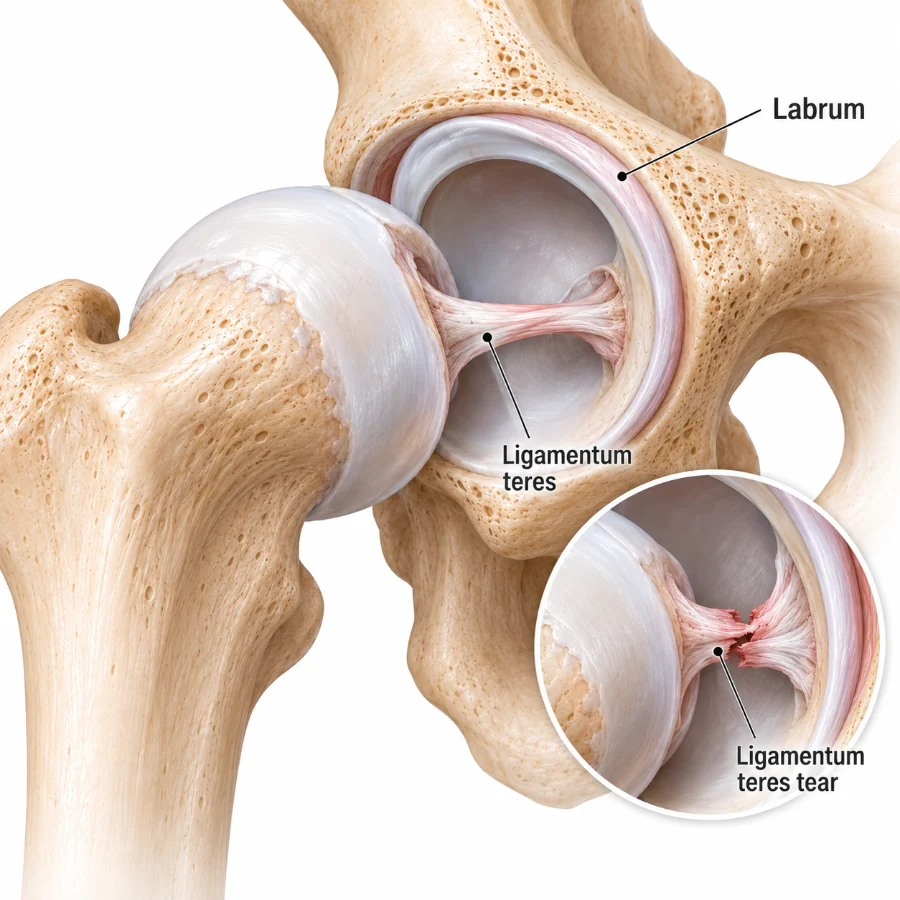
The ligamentum teres (LT) is a robust, cord-like structure inside the hip that connects the femoral head to the acetabulum. Traditionally thought to be a vestigial ligament, modern research shows the LT contributes to hip stability, particularly near the end ranges of rotation and flexion. It also contains pain fibres, which is why LT injuries can produce deep groin pain, catching, and a sense of giving way.

  • Primary role: dynamic restraint and proprioceptive feedback, supporting the capsule and surrounding muscles.
  • Clinical relevance: tears may coexist with femoroacetabular impingement (FAI), capsular laxity, or borderline dysplasia, and can be a hidden cause of persistent symptoms.

Symptoms of a Ligamentum Teres Tear

Symptoms range from vague discomfort to activity-limiting instability. Common features include:

  • Deep groin pain often described with the “C-sign,” sometimes radiating to the lateral hip or buttock.
  • Clicking, catching, or locking with twisting, pivoting, or getting in/out of the car.
  • Instability sensations or the hip feeling “untrustworthy,” especially during cutting, hopping, or rapid direction changes.
  • Worse with end-range movements such as deep flexion, combined rotation, or splits.
  • Possible night discomfort when rolling in bed or after high-load sessions.

Symptoms can overlap with a labral tear, chondral injuries, or adductor/psoas issues, so a careful assessment is important.

Common Causes & Risk Factors

  • Trauma: a forced twist, slip, tackle, or awkward landing can cause partial or complete tearing.
  • Repetitive microtrauma: high-load rotational sports (football, AFL, soccer, dance, martial arts) can fray the ligament progressively.
  • FAI morphology: cam or pincer shapes increase shear and impingement, placing stress on the LT and labrum.
  • Generalised laxity or capsular insufficiency: hypermobility syndromes predispose to micro-instability and LT injury.
  • Borderline dysplasia or acetabular under-coverage: the LT may compensate for limited bony stability and become overloaded.
  • Degenerative change: attritional tears can coexist with cartilage wear in older or highly active patients.

How We Diagnose a Ligamentum Teres Tear

Diagnosis combines a detailed history, targeted examination, and selective imaging. Because LT tears often coexist with other pathology, we assess the whole hip to identify all contributors to pain and instability.

Clinical Examination

  • Provocative tests: flexion-adduction-internal rotation (FADIR), scour/rotation tests, and instability/apprehension manoeuvres.
  • Functional control: single-leg squat, lumbopelvic stability, gluteal activation, foot mechanics, and core control.
  • Range of motion: looking for painful end-range rotation or asymmetry compared with the opposite side.

Imaging

  • X-rays: assess morphology (cam/pincer), version, and joint space to screen for arthritis or dysplasia.
  • MRI or MR arthrogram: may show LT signal change, partial tears, or synovitis; sensitivity varies and a normal scan does not exclude a tear.
  • CT (selected cases): defines bone anatomy when planning surgery for coexistent FAI or dysplasia.
  • Diagnostic injection: image-guided local anaesthetic can confirm the hip joint as the main pain source and assist in planning.

In some patients, the definitive diagnosis is made during hip arthroscopy, where the LT can be visualised directly and probed for stability.

Non-Operative Treatment

Many patients respond well to a structured, criteria-based rehabilitation program. Our physiotherapy team focuses on pain reduction, stability restoration, and graded return to load.

1) Education & Load Management

  • Modify or reduce end-range rotation and deep flexion that trigger pain or instability.
  • Temporarily down-scale high-impact or cutting activities; introduce cross-training (bike, pool, elliptical).
  • Optimise sitting hygiene (breaks every 30–45 minutes); adjust car and desk ergonomics.

2) Physiotherapy & Exercise Therapy

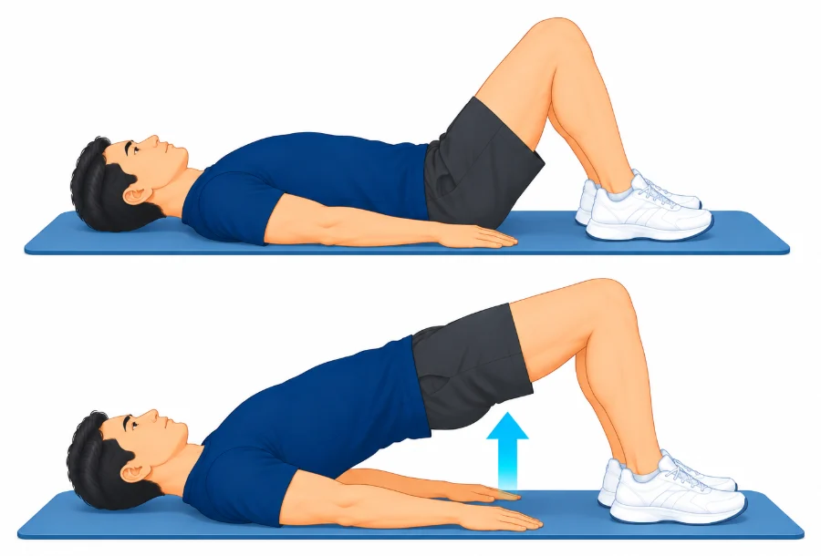
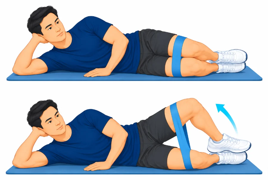
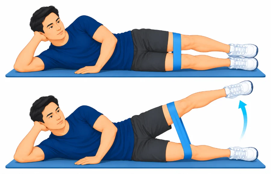
  • Gluteal strengthening (medius/maximus) for frontal-plane control.
  • Deep external rotators activation (short rotators) to stabilise the femoral head.
  • Core and lumbopelvic control to manage torsional loads through the hip.
  • Progressive proprioception: single-leg stance, anti-rotation work, perturbation training.
  • Gradual return-to-run and change-of-direction programs once criteria are met.

3) Medications & Injections

  • Short, targeted courses of anti-inflammatory medication if appropriate.
  • Image-guided corticosteroid injections may calm synovitis and enable rehabilitation.
  • Platelet-Rich Plasma (PRP) can be considered in selected cases after discussion of current evidence and goals.

Expected timeline: With consistent rehabilitation, many patients experience meaningful improvement within 6–12 weeks. Persistent mechanical symptoms (catching/instability) or failure to progress despite quality rehab may prompt a discussion about surgery.

When Is Surgery Considered?

We consider hip arthroscopy for confirmed or strongly suspected LT tears when best-practice non-operative care has not restored function, or when examination and imaging show associated mechanical problems such as significant FAI, capsular laxity, or borderline dysplasia that are unlikely to settle without operative correction.

Arthroscopic Treatment Options

  • Debridement (partial tears or fraying): removal of unstable fibres can reduce mechanical catching and pain.
  • Ligamentum teres repair: suture repair is considered in focal, repairable tears with good tissue quality.
  • Ligamentum teres reconstruction: in irreparable tears with symptomatic instability, a graft (e.g., hamstring or allograft) may be used to recreate restraint.
  • Capsular closure or plication: restoring capsular tension helps stability, especially in laxity or micro-instability.
  • Concomitant procedures: address FAI (cam/rim work) and any labral tear to reduce recurrent shear forces.

Surgery is tailored to the individual’s anatomy, tissue quality, and goals. We will outline the benefits, risks, and alternatives to help you make an informed decision.

Risks & Considerations

  • Infection, bleeding, clots (rare), temporary nerve irritation, stiffness.
  • Persistent pain if unrecognised co-pathology (e.g., significant cartilage loss or instability) remains.
  • Need for further surgery if symptoms recur or underlying morphology is not fully corrected.

Learn more about the procedure and preparation at Hip Arthroscopy (Keyhole Surgery).

Recovery & Rehabilitation

Rehabilitation is guided by the specific procedure(s) performed and your baseline strength and control. A typical pathway after arthroscopy:

  1. Week 0–2: protected weight-bearing as advised; crutches initially; swelling control; gentle range; isometrics (gluteals, deep rotators); avoid end-range rotation.
  2. Week 3–6: progress weight-bearing; introduce stationary bike and pool work; restore gait pattern and pelvic control; begin light proprioception.
  3. Week 6–12: advance strengthening (gluteals, rotators, core); controlled anti-rotation and perturbation drills; graded return to jogging once criteria are met.
  4. Month 3–6: sport-specific change-of-direction, acceleration/deceleration, and contact-prep as appropriate; staged return to training then competition.

Desk-based roles often resume within 1–2 weeks with ergonomic adjustments. Manual or pivoting sports require a longer, criteria-based progression monitored by our physiotherapy team.

Outcomes: What to Expect

When coexisting issues are addressed (FAI, capsular laxity, labral pathology) and rehabilitation is followed closely, many patients experience meaningful improvements in pain, stability, and function. Return-to-sport rates are favourable in well-selected cases—especially when objective strength and control milestones guide each stage of progression.

Factors associated with better outcomes include early treatment before extensive cartilage damage, appropriate capsular management, and adherence to a progressive, criteria-based program. If imaging shows established arthritis with joint-space narrowing, arthroscopy is less predictable; in these scenarios we will discuss whether muscle-sparing hip replacement offers a more reliable outcome.

Prevention & Performance Tips

  • Build gluteal strength and deep hip rotator control to support joint stability.
  • Periodise training loads; avoid sudden spikes in volume, intensity, or end-range rotation.
  • Technique coaching for cutting, kicking, and landing to limit excessive torsional stress at the hip.
  • Use cross-training (bike or pool) during flare-ups to maintain fitness while symptoms settle.
  • Address foot and trunk mechanics to reduce compensatory torque on the hip.

When Should I See a Hip Specialist?

Seek an expert review if you experience groin pain with catching, a sensation of instability, or symptoms that persist beyond a few weeks despite rest and activity modification. Early assessment helps confirm the diagnosis, rule out co-pathology, and start a targeted, effective plan.

Ligamentum Teres Tear — FAQs

Is a ligamentum teres tear the same as a labral tear?

No. The ligamentum teres is an intra-articular ligament, while the labrum is a fibrocartilage rim around the socket. They can be injured together and produce similar symptoms.

Can a ligamentum teres tear heal without surgery?

Many patients improve with load modification and targeted physiotherapy. Persistent instability or mechanical symptoms despite quality rehab may warrant arthroscopic treatment.

How long is recovery after arthroscopic treatment?

Desk work often resumes in 1–2 weeks. Jogging commonly begins around 8–12 weeks if criteria are met. Return to pivoting sport is typically within 3–6 months, guided by objective milestones.

Do you always reconstruct the ligamentum teres?

No. Debridement or repair may be sufficient in many cases. Reconstruction is reserved for irreparable tears with symptomatic instability, typically alongside capsular management and correction of bony conflict.

When is hip replacement a better option?

When imaging shows advanced arthritis with joint-space loss, arthroscopy outcomes are less predictable. In such cases, a modern muscle-sparing hip replacement may offer a more reliable outcome.