
Femoroacetabular impingement (FAI)
Femoroacetabular impingement, often called FAI, happens when the shape of the hip leads to extra contact between the ball and socket as the hip moves. It may cause groin pain with sitting, bending, twisting or sport. Many people improve with activity changes and a targeted rehabilitation program.
At the Specialist Orthopaedic Surgery Clinic in East Melbourne, orthopaedic assessment, physiotherapy, myotherapy and strength testing are all available in the same rooms, so the plan can be built around your hip and the activities you want to get back to.

What is femoroacetabular impingement?
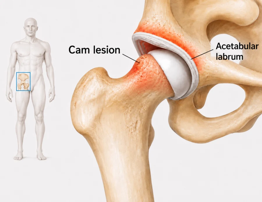
The hip is a ball and socket joint. In FAI the ball, the socket or both have a slightly different shape, so the bones meet earlier than usual when the hip bends or turns. A bump on the ball is called a cam shape, and extra cover from the socket rim is called a pincer shape.
Repeated contact may irritate the labrum, the ring of firm cartilage around the socket, or the smooth cartilage lining the joint. Many people have these hip shapes with no pain at all, so treatment is guided by your symptoms and examination and not by the scan alone.
Does this sound like you?
Arrange an assessment if groin or hip pain keeps returning, limits sport or exercise, reduces your hip movement, or has not improved with a suitable period of rehabilitation. Review is also useful when clicking or catching is painful.
How it is diagnosed
Diagnosis starts with the movements and positions that bring on pain and whether there is clicking, catching or lost movement. Hip movement and strength are tested, along with how the pelvis and leg move during tasks such as squatting and stepping.
MRI may be used to look at the labrum and cartilage when the result would change treatment. CT is occasionally helpful to map bone shape in detail before surgery, but it is not routinely needed.
Treatment and recovery
Treatment depends on whether the hip shape and examination match your symptoms, and on the health of the cartilage and labrum. Many people start with activity changes and a progressive physiotherapy program.
First weeks
Settle the hip
The positions that keep flaring the pain are identified and adjusted, such as deep squats or long periods in a low chair, while you stay active.
Physiotherapy
Build control
Exercises build hip and trunk strength and movement control. Myotherapy may ease tight muscles alongside the exercise program.
Building up
Return to activity
Load is increased gradually for sitting, running, sport and work. Strength testing can measure progress and guide return to higher level activity.
If progress stalls
Review the plan
If pain remains limiting despite a structured program, an injection or a surgical opinion may be discussed. Injections are not given at the clinic. If one is recommended, your GP can arrange it, usually through a radiology practice.
Exercises you may be given
Your physiotherapist will choose the right ones and the right level for you.
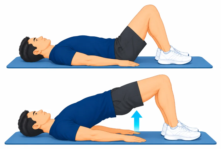
Bridge
Lie on your back with knees bent and feet flat. Squeeze your buttocks and lift your hips until your body is in a straight line, then lower slowly.
2 to 3 sets of 10
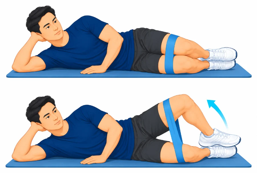
Clam
Lie on your good side with hips and knees bent. Keep your feet together and lift the top knee without rolling your pelvis back, then lower.
2 to 3 sets of 10 to 15
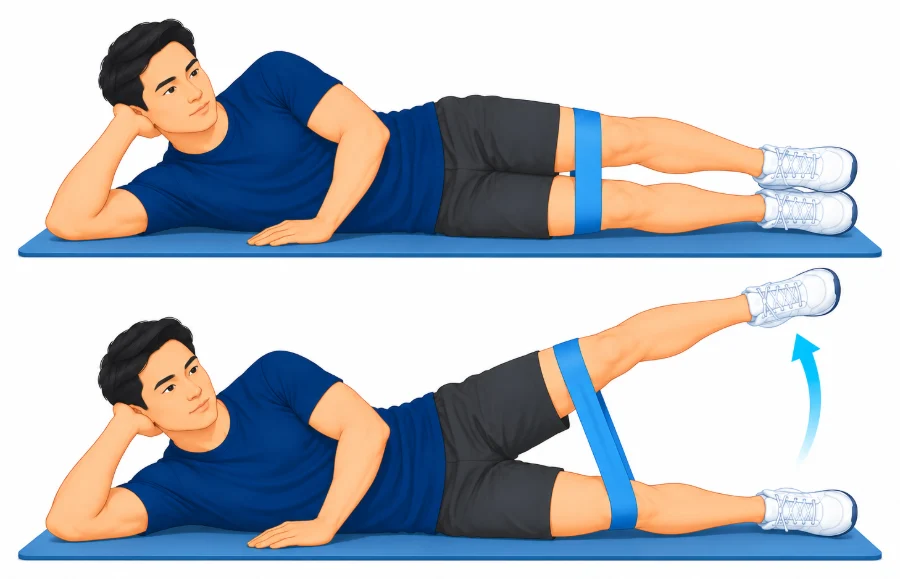
Side-lying leg lift
Lie on your good side with the lower knee bent. Keep the top leg straight and slightly behind you, lift it a short way, then lower slowly.
2 to 3 sets of 10
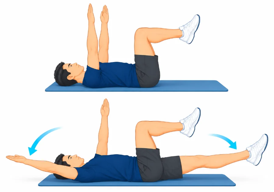
Dead bug
Lie on your back with hips and knees bent to 90 degrees and arms reaching up. Keep your lower back gently flat as you lower the opposite arm and leg, then swap.
2 sets of 8 each side
Other treatment options
Injections
An image-guided injection into the hip joint may give temporary relief and can sometimes help show whether pain is coming from inside the joint. It does not change the shape of the hip.
When surgery is needed
Hip arthroscopy may be considered when groin pain and loss of function remain significant despite a structured rehabilitation program, and examination and imaging show a treatable impingement problem without advanced arthritis. A cam or pincer shape seen on a scan is not a reason for surgery by itself. Prof Phong Tran performs hip arthroscopy, and his website explains the options and recovery.
Common questions
Does FAI always need surgery?
No. Many people improve with activity changes and targeted physiotherapy. Surgery is considered only when symptoms remain limiting, the examination and imaging match, and there is no advanced arthritis.
Can a cam or pincer shape exist without pain?
Yes. These hip shapes are common in people with no symptoms. Treatment is directed at your pain and function.
What exercises help hip impingement?
A program usually focuses on hip and trunk strength, movement control and a gradual return to your usual activities. Exercises are adjusted if deep hip bending causes a flare.
Will an injection confirm the diagnosis?
An image-guided local anaesthetic injection can sometimes help show whether pain is coming from inside the hip. It is one part of the assessment, alongside the history, examination and imaging.
How long is recovery after hip arthroscopy?
Crutches are commonly used for one to three weeks. Running and sport are introduced over several months, and return to higher demand activity often takes four to six months or longer.
Book an assessment
An assessment can clarify whether your hip symptoms are related to FAI and which treatment suits your activities and goals. Physiotherapy with A/Prof Amir Takla and Imogen Clark, myotherapy with Joanne VanDer Haar, and specialist review with Prof Phong Tran, all at 166 Gipps Street, East Melbourne. Please bring any previous hip imaging and details of rehabilitation or other treatment already tried.