
Baker’s cyst
A Baker’s cyst is a fluid-filled swelling at the back of the knee. It usually forms because the knee is making extra fluid, often from arthritis, a meniscus problem or inflammation inside the joint. Treatment mostly focuses on the knee problem underneath, and surgery on the cyst itself is rarely needed.
At the Specialist Orthopaedic Surgery Clinic in East Melbourne, orthopaedic assessment, physiotherapy and myotherapy all happen in the same rooms, so the swelling and the knee problem behind it can be looked at together.

What is a Baker’s cyst?
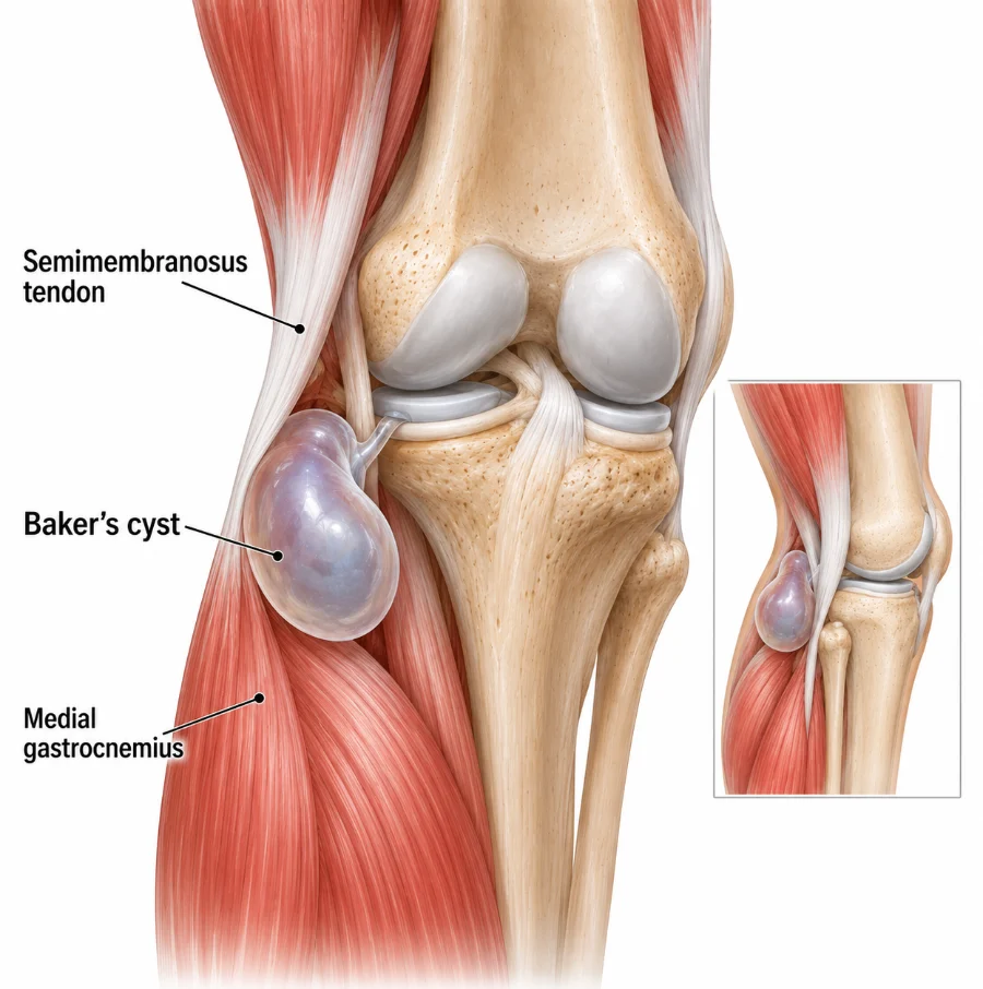
When the knee is irritated it can make more joint fluid than usual. Some of this fluid can pass through a small opening into a sac at the back of the knee, forming a soft swelling.
The cyst is usually a sign of another knee problem, such as osteoarthritis, a meniscus tear or joint inflammation. Its size can change over time, and some cysts cause no symptoms at all. It is not a tumour.
Does this sound like you?
Arrange an assessment if swelling behind the knee persists, becomes painful, limits movement or keeps coming back, especially with other knee symptoms such as arthritis pain or locking.
How it is diagnosed
Diagnosis starts with examining the swelling at the back of the knee and checking the knee for an underlying cause such as arthritis or a meniscus problem.
Ultrasound can confirm the swelling contains fluid and tell a cyst apart from a blood vessel problem or another lump. MRI is usually kept for uncertain cases or when another problem inside the knee needs to be checked. Sudden calf swelling may need urgent testing for a blood clot.
Treatment and recovery
Treatment usually targets the knee problem that is making extra fluid, since the cyst tends to settle when the joint settles. Most people are managed with activity changes, physiotherapy and treatment of the underlying condition.
First weeks
Settle the swelling
Activity is adjusted while the knee settles, without resting it completely. Compression may help comfort.
Physiotherapy
Restore movement
Gentle exercises regain knee bending and straightening and build thigh and hip strength.
Treat the cause
Manage the knee
Arthritis, a meniscus tear or joint inflammation is treated directly, which often reduces the fluid feeding the cyst.
If it keeps returning
Review the plan
Aspiration, an injection or further imaging may be arranged if the cyst stays painful or keeps coming back. Injections are not given at the clinic. If one is recommended, your GP can arrange it, usually through a radiology practice.
Exercises you may be given
Your physiotherapist will choose the right ones and the right level for you.
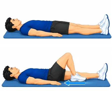
Heel slides
Lie on your back with legs straight. Slowly slide the heel of the sore leg towards you to bend the knee as far as is comfortable, then slide it back out.
10 slow slides, twice a day
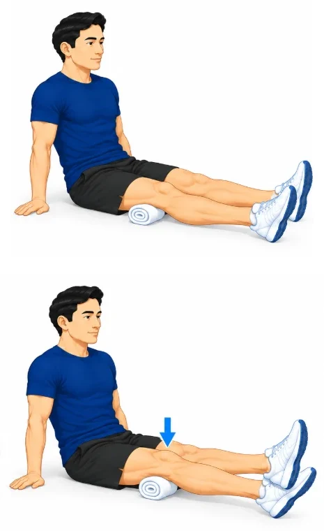
Quadriceps tightening
Sit with your leg straight. Tighten the thigh muscle to press the back of the knee gently down, hold, then relax.
10 holds of 5 seconds
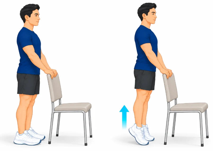
Calf raises
Stand holding the back of a chair for balance. Rise slowly onto your toes, then lower your heels back down.
2 sets of 10 to 15
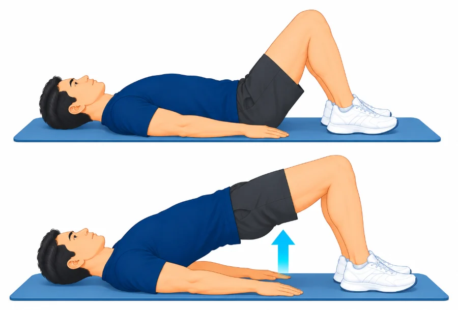
Bridge
Lie on your back with knees bent and feet flat. Squeeze your buttocks and lift your hips until your body is in a straight line, then lower slowly.
2 sets of 10
Other treatment options
Aspiration and injections
Ultrasound-guided draining of the cyst, or a corticosteroid injection into the knee, may give temporary relief in selected cases. The cyst may return if the underlying arthritis, inflammation or meniscus problem remains active.
When surgery is needed
Surgery on a Baker’s cyst is rarely needed. It may be considered when a large cyst stays painful or keeps returning despite treatment of the knee problem underneath, or presses on nearby nerves or blood vessels. Any surgery usually treats the underlying knee problem first, and a cyst can still return. Prof Phong Tran can review whether surgery is appropriate for you.
Common questions
Is a Baker’s cyst a tumour?
No. It is a fluid-filled swelling behind the knee, usually related to irritation inside the joint.
Why does the cyst keep coming back?
It can return while arthritis, inflammation or another knee problem keeps making extra joint fluid.
Can a Baker’s cyst burst?
Yes. Fluid can track into the calf and cause sudden pain and swelling, which can look like a blood clot and needs assessment.
Should the cyst be drained?
Drainage may give temporary relief in selected cases, but the cyst often returns unless the knee problem underneath is treated.
Is surgery usually required?
No. Treatment normally focuses on the underlying knee condition, and removing the cyst directly is uncommon.
Book an assessment
An assessment can find the knee problem behind the swelling and set out treatment that suits you. Physiotherapy with A/Prof Amir Takla and Imogen Clark, myotherapy with Joanne VanDer Haar, and specialist review with Prof Phong Tran, all at 166 Gipps Street, East Melbourne. Please bring any previous knee X-rays, ultrasound or MRI reports.