Calcific tendinitis of the shoulder
Calcific tendinitis happens when a calcium deposit forms inside one of the rotator cuff tendons. It may cause a sudden episode of severe shoulder pain or a slower ache with lifting and reaching. Many cases settle with non-operative treatment, and deposits often shrink or disappear over time.
At the Specialist Orthopaedic Surgery Clinic in East Melbourne, orthopaedic assessment, physiotherapy, myotherapy and review of your X-ray or ultrasound are brought together, so treatment can step up only as far as your shoulder needs.
What is calcific tendinitis?
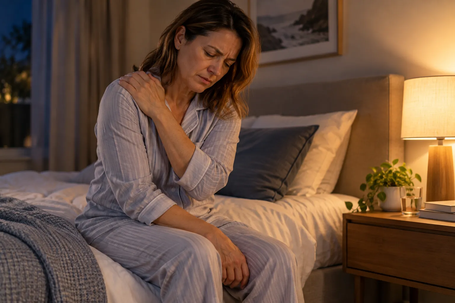
The rotator cuff is a group of tendons that lift and turn the arm. In calcific tendinitis a deposit of calcium forms within one of these tendons, most often the tendon across the top of the shoulder. The deposit lies within the tendon itself and is not part of the bone.
A deposit may stay quiet for a long time. When the body starts to break it down, the area can become inflamed and very painful, and the nearby bursa may also be irritated. The exact cause is not fully understood. It is not caused by eating too much calcium and does not mean your blood calcium is high.
Does this sound like you?
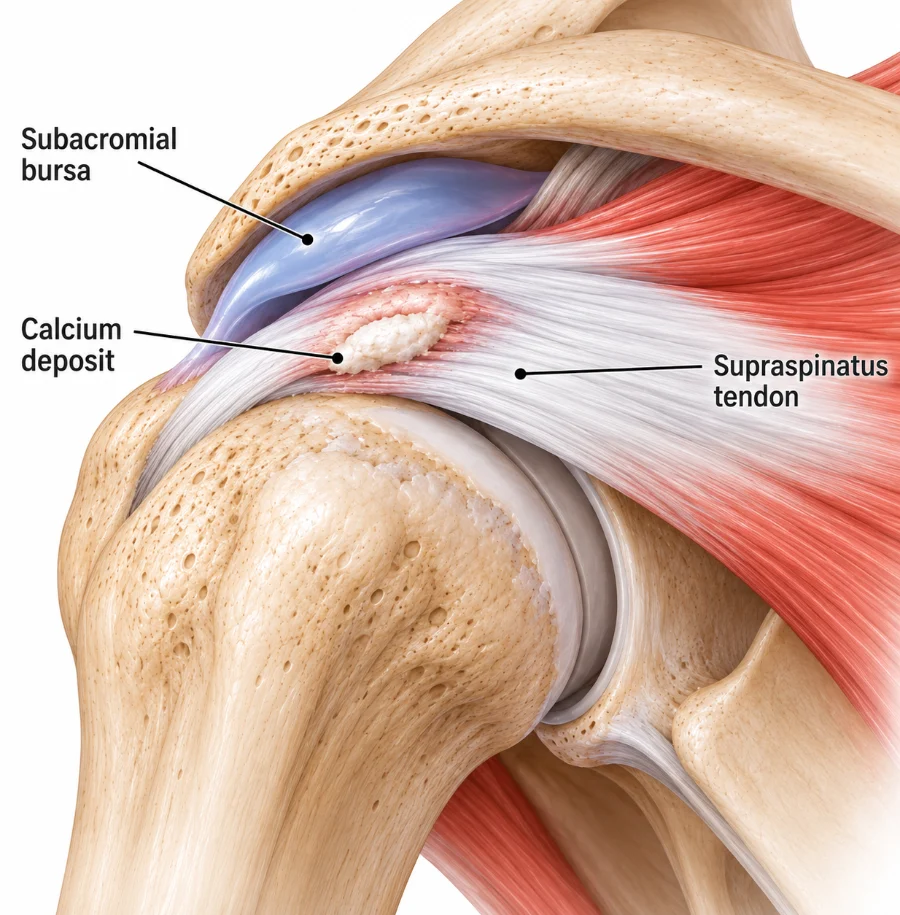
Arrange an assessment if shoulder pain persists, keeps returning, limits movement or sleep, or has not settled with initial treatment. A sudden severe flare may also need prompt medical review.
How it is diagnosed
Diagnosis starts with whether the pain came on suddenly or gradually, and an examination of shoulder movement and strength. The neck is also checked as another possible source of pain.
A standard X-ray often shows the calcium deposit clearly, along with its size and position. Ultrasound adds detail, checks the rotator cuff and can guide needling or an injection. MRI is not routinely needed.
Treatment and recovery
Treatment depends on how severe and long-standing the pain is and whether the deposit appears to be changing. Many episodes improve with pain relief, short-term activity changes and physiotherapy.
Acute flare
Settle the pain
Painful activity is reduced for a short time and pain relief is discussed with your GP or pharmacist. Gentle movement helps prevent stiffness.
Physiotherapy
Restore movement
As pain settles, exercises restore shoulder movement and then rebuild rotator cuff and shoulder blade strength.
If pain persists
Targeted treatment
An injection, ultrasound-guided barbotage or shockwave therapy may be considered, followed by further rehabilitation. Injections are not given at the clinic. If one is recommended, your GP can arrange it, usually through a radiology practice.
If still limited
Review the plan
If severe pain and loss of function continue despite these steps, a surgical opinion may be arranged.
Exercises you may be given
Your physiotherapist will choose the right ones and the right level for you.
Pendulum swings
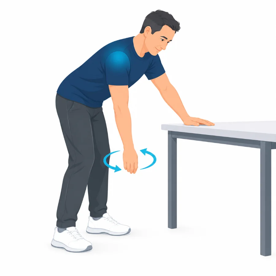
Lean on a bench and let the sore arm hang. Rock your body so the arm swings in small, relaxed circles.
30 seconds, a few times a day
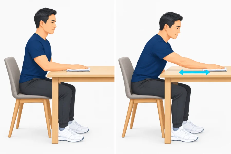
Table slides
Sit beside a table with your hand on a small towel. Slowly slide the hand forward as far as is comfortable, then back.
10 slow slides, twice a day
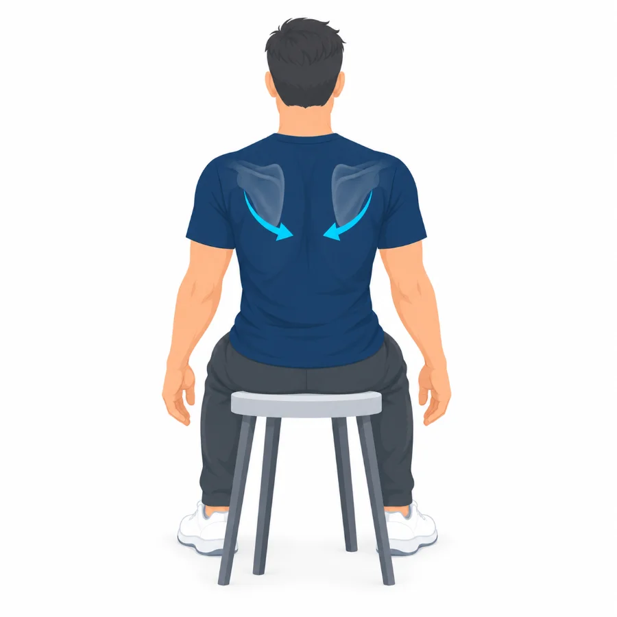
Shoulder blade squeeze
Sit tall and gently draw your shoulder blades back and down. Hold, then relax without shrugging.
10 holds of 5 seconds
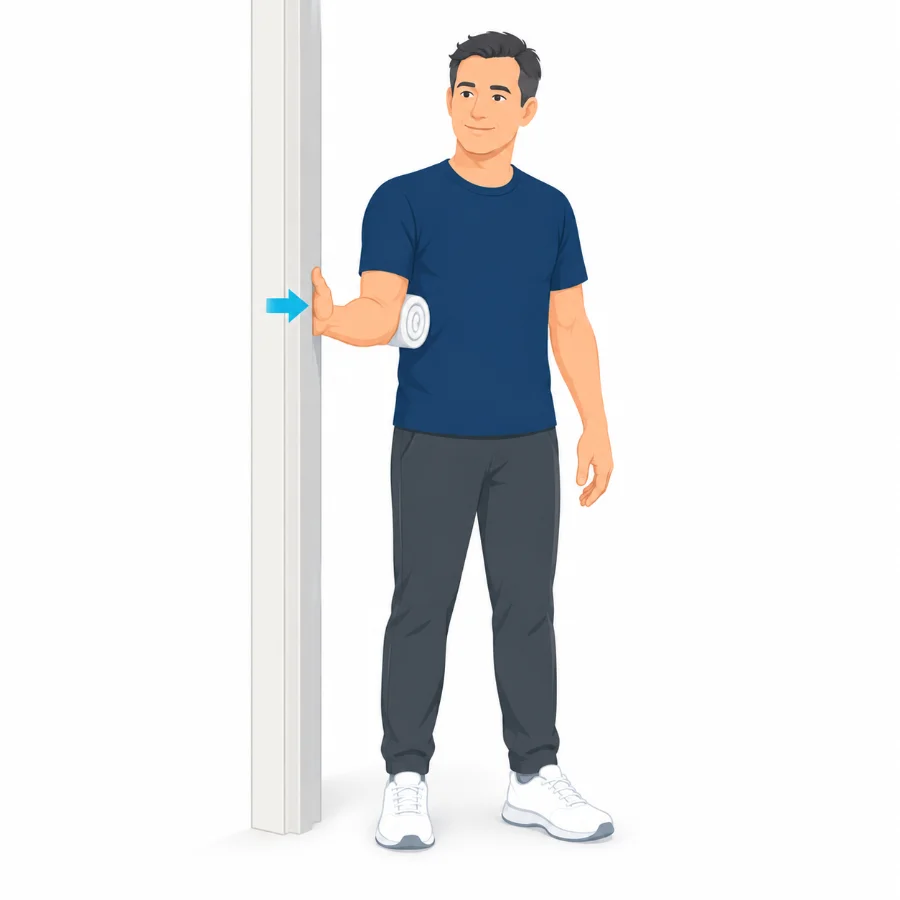
Gentle outward press
Stand side-on to a wall with your elbow bent and tucked in. Press the back of your hand gently into the wall without moving the arm.
5 to 10 presses, hold 5 seconds
Other treatment options
Injections, barbotage and shockwave
A corticosteroid injection into the bursa may reduce pain and inflammation. For a persistent deposit, ultrasound-guided barbotage uses a fine needle to break up and rinse out some of the calcium. Shockwave therapy may help in selected people. These are followed by rehabilitation.
When surgery is needed
Arthroscopic removal of the calcium deposit is uncommon. It may be considered when severe pain and loss of function persist despite rehabilitation, injection, barbotage or shockwave therapy, and the deposit clearly matches the symptoms. Prof Phong Tran performs shoulder arthroscopy, and his website explains the options and recovery.
Common questions
Is it caused by too much calcium in my diet?
No. It is not linked to eating calcium and does not mean your blood calcium level is high.
Will the deposit go away?
Many deposits shrink or disappear over time, sometimes after a painful flare as the body breaks them down.
Why is the pain so severe?
When the body starts to reabsorb the deposit, the tendon and nearby bursa can become very inflamed. This phase often settles.
What is barbotage?
An ultrasound-guided procedure where a fine needle is used to break up and rinse out some of the calcium, usually with local anaesthetic.
Do I need surgery?
Rarely. Surgery is kept for ongoing severe pain and loss of function after suitable non-operative treatment.
Book an assessment
An assessment can review the deposit and set out treatment options that suit your symptoms. Physiotherapy with A/Prof Amir Takla and Imogen Clark, myotherapy with Joanne VanDer Haar, and specialist review with Prof Phong Tran, all at 166 Gipps Street, East Melbourne. Please bring any previous X-rays, ultrasound reports and details of treatment already tried.